Chapter 1 - Introduction
Ultrastructure is the architecture of cells that is visible at higher magnifications than found on a standard optical light microscope. Electron microscopes is widely used to investigate the ultrastructure of biological specimens.
This chapter demonstrates the potential of electron microscopy in understanding histology.
 |
Transmission Electron Microscopy (TEM)
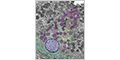
TEM produces two-dimensional images of a specimen by imaging a thin section with a beam of electrons. Ultrathin tissue sections are stained with heavy metals (such as osmium tetroxide, uranium or lead salts). The images are colorless and have a resolution of ~0.1 nm (~1000 times greater than a light microscope).
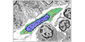
Example micrograph of a cell imaged by TEM that explains how these images should be viewed.
 |
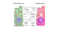
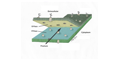
Schematic comparison of a cell imaged by TEM and light microscopy.
 |
Figures
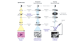
Schematic representations of a simple columnar cell from images acquired by TEM, SEM, and freeze fracture.
 |
|
 |
|
 |
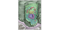
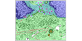
Ultrastructure of Cells (TEM)
 |
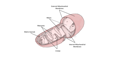
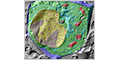
(Nucleus / Golgi Apparatus / Mitochondria / Endoplasmic Reticulum) |
 |
|
 |
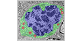
(Nucleus / Centrosome / Microtubules) |
 |
|
 |
|
 |
|
 |
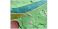
Scanning Electron Microscopy (SEM)
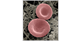
Scanning electron microscope (SEM) produces three-dimensional images of a speciment by measuring the relative differences in the reflection of a focused beam of electrons scanned across a specimen. Biological specimens are usually coated with a thin layer of metal (such as platinum) to form a replica that is then imaged. This allows the surface structures of organelles, cells, and tissues to be visualized. The images are colorless and have a resolution of ~10 nm (~100 times greater than a light microscope).
 |
|
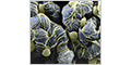 |
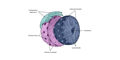
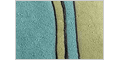
Freeze Fracture
Freeze fracture technique reveals the internal structure of biological specimens. Frozen specimens are physically broke apart (fracturing) and the exposed surface is coated with evaporated platinum. This replica is then imaged by TEM.
 |
Freeze fracture forms enface views of membrane-bound compartments that give striking three-dimensional representations.
 |
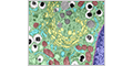
Ultrathin Section versus Freeze Fracture
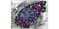
Mast cell as seen in an ultrathin sections and by freeze fracture.
 |
|
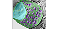 |
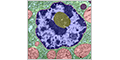
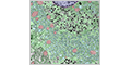
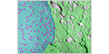
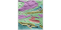
Ultrastructure of Cells (Freeze Fracture)
Freeze fracture reveals interior surfaces of cells.
 |
|
 |
|
 |
|
 |
|
 |
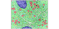
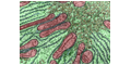
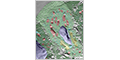
Mitosis
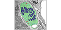
Cells undergoing mitosis as seen by TEM.
 |
|
 |