Chapter 1 - Introduction
The goal of this chapter is to learn how to look for items of interest in histological specimens using light microscopy. A variety of cells, tissues, and organs are provided as samples.
Partial list of characteristics to notice and observe:
- Size of cells
- Shape of cells
- Nuclear/cytoplasmic ratio
- Staining properties
- Basophilic or acidophilic
- Heterochromatin and euchromatin
- Special staining properties
Histological Stains
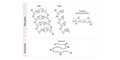
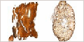
Biological material is inherently of low contrast and provides little to see in a standard bright field microscope unless treated with a histological stain. Hematoxylin and eosin are the most widely used dyes in histology and pathology. The following slides demonstrate the staining characteristics of these dyes alone, and more importantly, in combination.
 |
|
 |
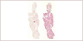
Cells and Tissues
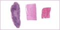
The cell is the basic structural and functional unit of all living organisms. Cells vary widely in size and shape depending on their function. Microscopes are used to study cells because most cannot be seen with an unaided eye.
It is not necessary to learn the names of specific cells and tissues for this chapter, but rather learn to recognize variations in the size, shape, and staining properties of cells.
 |
|
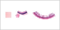 |
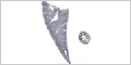
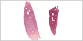
Other Stains
 |
(Nissl Substance / Metachromasia) |
 |
(Golgi method) |
 |
(Iron hematoxylin) |
 |
(Feulgen reagent) |
 |
(Periodic acid-Schiff reagent) |
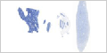
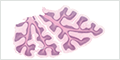
Purkinje Cells
Different stains used to visualize Purkinje cells in the cerebellum.
 |