Chapter 3 - Connective Tissue
Connective tissue supports and protects organs. It has three primary components: fibers, extracellular matrix and cells. The composition of these elements is the basis of which connective tissues are classified.
This chapter examines the basic types of connective tissue, while subsequent chapters examine specialized connective tissues such as cartilage, bone and blood.
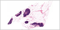
Mesenchyme
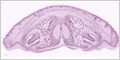
Mesenchyme is an early form of connective tissue found in embryos. Mesenchymal cells can differentiate into other cell types such as fibroblasts, fat cells, cartilage and bone.
 |
|
 |
Connective Tissue Fibers
Connective tissue contains three types of fibers: collagen, elastic and reticular.
 |
(H&E / azan / Verhoeff) |
 |
(silver) |
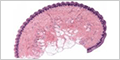
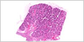
TYPES OF CONNECTIVE TISSUE
Connective tissue is classified based on the density of fibers (loose or dense), the arrangement of fibers (irregular or regular) and the predominate fiber or cell type.
 |
MH 021-022-023 Connective Tissue (H&E / azan / Verhoeff) |
Loose Connective Tissue
Loose (areolar) connective tissue has a sparse and random arrangement of fibers.
|
|
 |
Dense Regular Connective Tissue
Dense regular connective tissue contains many collagen fibers arranged in parallel bundles.
 |
Dense Irregular Connective Tissue
Dense irregular connective tissue contains collagen fibers that are more randomly arranged.
 |
CELLS OF CONNECTIVE TISSUE
Although connective tissue has fewer cells than most tissues, the cells found in connective tissue are still important. Fibroblasts and adipocytes do not leave connective tissue.
Fibroblast
Fibroblasts are widely distributed within connective tissue and synthesize the components of the extracellular matrix. They are also capable of differentiating into other types of connective tissue cells.
|
|
|
|
 |
Adipocyte
Adipocytes (or fat cells) are specialized for the synthesis and storage of lipids. They may occur singly but are more often found as clusters within loose connective tissue.
|
|
 |
(mesentery) |
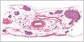
TRANSIENT CELLS OF CONNECTIVE TISSUE
Neutrophils, eosinophils, basophils, monocytes, lymphocytes, plasma cells and mast cells are immune cells that migrate from the blood into connective tissues.
Eosinophil
Eosinophils are involved in many inflammatory processes, including parasitic infections, allergic diseases, and asthma.
 |
Mast Cell
Mast cells are widely distributed in connective tissue. They release molecules that dilate blood vessels and recruit more immune cells to the site of an infection.
 |
(aldehyde fuchsin) |
 |
(mesentery) |
Plasma Cell
Plasma cells produce large quantities antibodies against specific antigens.
 |
(small intestine) |
Macrophage
Monocytes differentiate into macrophages within tissues. Macrophages are avidly phagocytic cells that engulf and digest microbes, cellular debris and foreign substances.
|